May 10, 2016
Optimizing the Ocular Surface Prior to Surgery: Why I Added Prokera to My Refractive Cataract Pre-Surgical Regimen
published on May 10, 2016 by
Refractive cataract surgery requires a high level of attention to the ocular surface. Patients expect perfect outcomes, and any compromise of the ocular surface interferes with preoperative measurements and proper IOL selection. To ensure we don’t end up with a postoperative refractive surprise, we need to be on alert for ocular surface disease and get it under control before proceeding with surgery.
Be Proactive
Our typical cataract surgery patients have all the risk factors for ocular surface problems. They have chronic disease comorbidities, they’re using multiple medications, and so forth. Many are neurotrophic to some degree; therefore, we can’t rely on them to report symptoms. We must proactively look for dry eye as well as epithelial-based diseases, such as Salzmann’s nodular degeneration (SND) and epithelial basement membrane dystrophy (EBMD). EBMD not only leads to irregularities in preoperative measurements and outcomes but also puts patients at risk for epithelial sloughing during surgery and poor wound healing postoperatively. Irregular topography, including irregular astigmatism, and discrepancies between K values obtained via biometry and topography are signs of EBMD. Also, a discrepancy between MMP-9 testing and tear osmolarity testing, perhaps one is positive and one is negative, is also a signal to look deeper for an underlying condition. For example, lifting the eyelid can reveal an EBMD ridge or conjunctivochalasis.
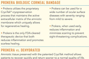
I don’t perform refractive cataract surgery for any patient who has dry eye or EBMD unless I can restore the health of the ocular surface first. For patients with dry eye and staining that persists after basic therapies, such as artificial tears or topical steroids, I use Prokera® Slim, a cryopreserved amniotic membrane (AM) fastened to a polycarbonate ring that is easily inserted into the patient’s eye in the clinic. The device is usually well tolerated and after approximately 1 week, I’m typically able to obtain accurate topography and biometry and proceed with surgery.
My solution for EBMD prior to refractive cataract surgery is superficial keratectomy followed by placement of Prokera Slim the next day. I remove the device in approximately 1 week and wait about 6 more weeks before repeating the cataract surgery measurements.

Pre-surgical Regimen
The ability of Prokera Slim to quickly normalize the ocular surface in chronic dry eye patients and accelerate re-epithelialization after superficial keratectomy has made it a very important part of my pre-cataract surgical regimen. I’ve retrospectively evaluated my outcomes, and it’s clear that Prokera Slim makes a significant difference for the better. I’m happy to report that I’ll soon be conducting a prospective clinical study to formally evaluate the use of Prokera Slim following superficial keratectomy for EBMD.
Elizabeth Yeu, MD is assistant professor of ophthalmology at Eastern Virginia Medical School and in private practice at Virginia Eye Consultants in Norfolk, Va.
