Promote Enhanced Ocular Healing by Incorporating Cryopreserved Amniotic Membrane Products In Your Treatment Protocol
The Gold Standard for Ocular Surface Reconstruction
The Gold Standard for Ocular Surface Reconstruction
Learn how it can help restore a healthy ocular surface for your patients by filling out the form and downloading a FREE AmnioGuard information guide.
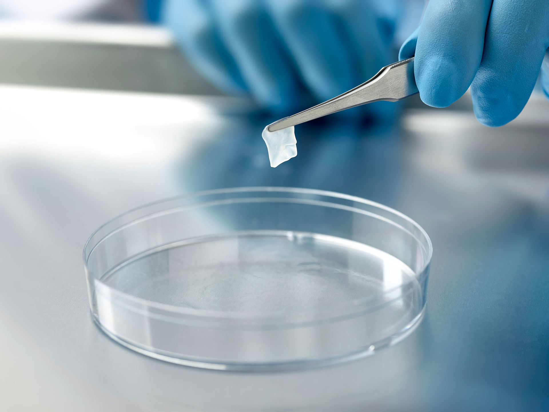
AmnioGuard is an umbilical cord graft used by ophthalmic surgeons for a number of indications including to cover the tube shunt of a glaucoma drainage device. Due to its high tensile strength and optimally designed thickness, AmnioGuard can reduce surgical times because it does not require rehydration, is easy to handle and suture, and can be used for corneal, conjunctival, sclera, fornix, socket and eyelids where thickness and high tensile strength are desired.
Ophthalmologists need proven outcomes.
The platform technology behind AmnioGraft® is supported by more than
500K
TRANSPLANTS
360
CLINICAL PUBLICATIONS
30Y
RESEARCH & DEVELOPMENT
Why AmnioGuard?
Ideal for Surgeries Requiring a Thicker Tissue Replacement
Created using the Company’s proprietary CryoTek® cryopreservation method, AmnioGuard delivers the natural properties of the amniotic membrane to help reduce inflammation, scarring and promote regenerative healing. AmnioGuard is a resilient and single-layer tissue which provides strong tectonic support for treating corneal, scleral, socket, fornix, and eyelid problems. For example, when it is used to cover the glaucoma shunt tube, it also allows visualization of the shunt tube and laser suture lysis and better cosmesis.
Reduce Inflammation, Promote Regenerative Healing
AmnioGuard is minimally manipulated tissue, which contains naturally-occurring biological factors that have been recognized as suppressing inflammation, preventing scar, and promoting ocular surface healing. The cryopreserved umbilical cord was developed to cover a wide variety of drainage devices to ensure superior surgical outcomes. AmnioGuard exerts anti-inflammatory and anti-scarring actions and works synergistically with a patient’s own system to enhance and speed tissue repair, resulting in a calm, white eye with minimal discomfort. AmnioGuard was developed specifically for tissues such as cornea, sclera, socket/fornix and eyelids where replacement with a thicker and stronger tissue is desired.
Why Intervene with AmnioGraft?
Discover AmnioGraft®
AmnioGraft® serves as a tissue replacement that also delivers the unique therapeutics actions of cryopreserved amniotic membrane. It contains the only amniotic membrane tissue with high tensile strength that retains intraoperative resilience and workability8 and ensures reproducible surgical outcomes.

Support Accelerated Post-Op Recovery1
AmnioGraft® expedites patient recovery by reducing inflammation5,9-11 and promoting fast, regenerative healing – typically within 2-3 weeks1-7. It helps prevents disease recurrence 1,2,6-8 as demonstrated in a study of 535 patients. AmnioGraft creates a durable recurrence barrier proven to support sustained healing with ≤5.8%* recurrence after 1 year1.
Creating a Healing Environment
AmnioGraft contains the only cryopreserved amniotic membrane that is FDA-cleared and designated for anti-inflammation, anti-scarring and anti-angiogenesis. Proprietary CryoTek preservation method retains the biologic properties and structural integrity is equivalent to fresh tissue2,12.
See What Others Are Saying About AmnioGraft
See What Others Are Saying About AmnioGraft
Mark Milner, MD
“Dry eye disease isn’t just simply a problem with quantitative tear film, meaning a low tear volume. Dry eye can also be from an abnormal tear film where you have tears, but they’re either unhealthy, or they’re not getting to the location that they need to, and one of the classic examples is what we call ‘Conjunctivochalasis’ or Mechanical Dry Eye.”
Neel Desai, MD
“We’ve all had the paradoxical patient that seems to have dry eye and typical ocular surface disease, but they simply don’t respond to all of the typical conventional therapies…there is a missing x-factor. And that x-factor is this Mechanical Dry Eye concept, or conjunctivochalasis.”
Cliff Salanger, MD
“Mechanical Dry Eye is rampant; it’s almost ubiquitous in individuals over the Medicare age of 65, and we have to look for it. The key is making the diagnosis. I get fooled still. I look, white light shining the slit beam across, and I’m thinking to myself, ‘Oh, there’s not a whole lot of redundant conjunctiva here.’ I put the fluorescein in, I turn on the cobalt blue light, and then even better, I put a yellow filter on the other side of the microscope, and it highlights the redundant fold of the conjunctiva.”
DISCLAIMER
BioTissue, Inc. will use reasonable efforts to include accurate and up-to-date information on this Website but makes no warranties or representations of any kind as to its accuracy, currency, or completeness.
The contents of this web site are protected by applicable copyright laws. No permission is granted to copy, distribute, modify, post or frame any text, graphics, video, audio, software code, or user interface design or logos.
The information contained on this website is for general information and educational purposes only, and is not intended to diagnose, treat, or cure any medical conditions. Please consult with your Physician for medical advice.
BioTissue, Inc. and its affiliates furnish this allograft product without any express or implied warranties. All statements or descriptions are informational only and are not to be interpreted or implied as a warranty of the allograft product. BioTissue, Inc. and its affiliates make no guarantee regarding the biological characteristics of this product. The end user shall be held responsible for determining the appropriate application and usage of this product.
